What to Expect During an Echocardiogram
A physician may recommend a variety of tests to assess the health of your heart. One common diagnostic tool is an echocardiogram; this procedure provides detailed images of the heart’s structures and function. Understanding what an echocardiogram is and what the procedure involves can help you prepare for your appointment, providing you more clarity throughout the process.
What Is an Echocardiogram?
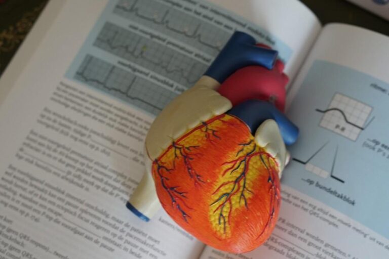
An echocardiogram is a non-invasive imaging test that allows a medical professional to observe the heart as it beats and pumps blood. It does this by using sound waves to create live images of the heart. These images can help a doctor evaluate the size, shape, and movement of the heart muscle, chambers, and valves. The test is performed by a sonographer, using a small, handheld device called a transducer.
There are several types of echocardiograms, and each serves a specific purpose. The most common type is the transthoracic echocardiogram (TTE), where the transducer is moved across the chest. Other types include the transesophageal echocardiogram (TEE), where a probe is guided down the throat. A stress echocardiogram is performed before and after physical activity to monitor the heart’s response to exertion. Your doctor will determine the most appropriate type of test based on your specific medical situation.
What Conditions Can It Detect?
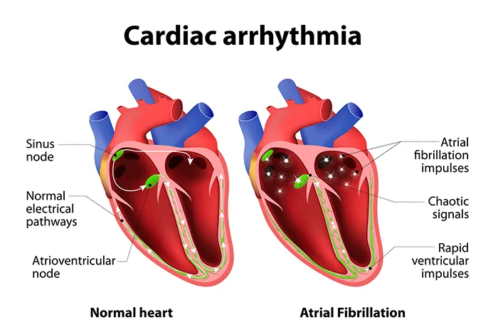
An echocardiogram provides a comprehensive view of the heart’s anatomy and function. This makes it a valuable tool for identifying a wide range of cardiac conditions. Echocardiograms can assess the overall size and shape of the heart. An enlarged heart or thickened heart walls may be signs of conditions like high blood pressure or heart failure. It can show how well the valves are operating, detecting problems such as a valve narrowing or leaking. The imaging can detect structural abnormalities. This includes congenital heart defects, which are problems with the heart’s structure present at birth. An echocardiogram can identify blood clots within the heart’s chambers, tumors, and problems with the sac that surrounds the heart.
What Should You Expect?
When you arrive for your appointment, you will be asked to undress from the waist up and put on a hospital gown. The sonographer will ask you to lie on an examination table. Small, sticky patches called electrodes will be attached to your chest, and are connected to an electrocardiograph (ECG) monitor that records your heart’s electrical activity during the test.
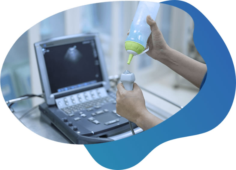
The sonographer may apply a special gel to your chest, helping the sound waves travel from the transducer to your heart. The sonographer will then press the transducer firmly against your skin and move it across your chest to capture images from different angles. You may be asked to change positions or hold your breath for short periods to help obtain clearer pictures.
Confer With a Heart Specialist
Following the echocardiogram, a cardiologist will review the images. The cardiologist will analyze the recordings to assess your heart’s structure and function and prepare a detailed report. Your doctor will then discuss the results with you at a follow-up appointment. The findings from the echocardiogram will help your doctor make a diagnosis, determine the severity of a condition, or guide the next steps in your treatment plan.