
Exploring the Neuroscience Behind Brain Surgery
Brain surgery addresses specific physical abnormalities located deep within the patient’s delicate brain tissue structures. Because the organ controls all bodily functions, surgeons operate with extreme and calculated precision. The skull provides strong protection, yet accessing the brain requires specialized medical instruments. Here is more information about the neuroscience behind brain surgery:
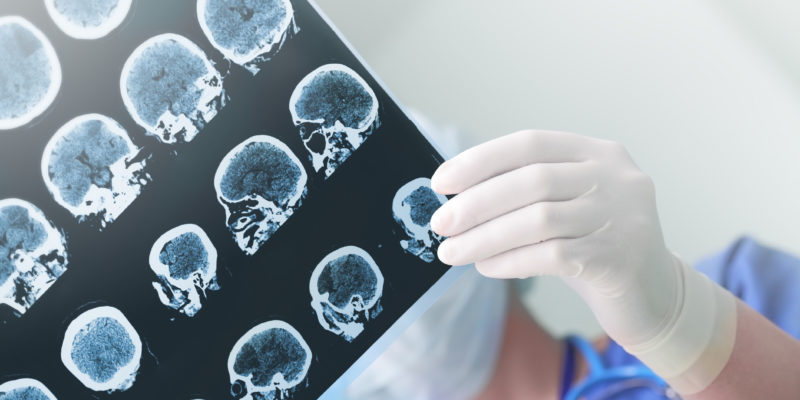
Advanced Imaging Techniques
Magnetic resonance imaging provides clear views, and CT scans show dense bone structures. Before the operation begins, these scans create a detailed 3D roadmap for surgical guidance. High-resolution images reveal the exact location of the targeted structural abnormality within the brain. Doctors review these images, and they plan the safest approach for entry and treatment.
Real-time imaging guides the surgeon’s hands during the actual complex medical procedure. The team monitors progress on screens, and adjustments happen in the exact moment. Because technology improves accuracy, surgeons avoid areas that control speech or vision functions. Intraoperative scans update the map as the brain shifts during the surgical process.
Direct Observation
While scans are useful, direct visual inspection remains a primary surgical tool for doctors. High-powered microscopes illuminate the operating field with intense, focused, bright light for clarity. Surgeons identify tissue texture changes, and they distinguish tumors from healthy cells visually. When the surgeon views the tissue, they verify what the scans suggested previously.
Neural Connectivity
When surgeons plan a procedure, they study the brain’s complex internal communication pathways. Nerves carry signals rapidly, and disrupting them affects basic movement or sensation in the body. Every neural connection serves a specific purpose for daily human functionality and interaction. The brain forms a vast network, and surgeons navigate this web carefully during the operation.
Mapping these areas prevents damage to healthy and active neural tissue near the site. Electrical signals travel constantly, so monitoring devices track this activity during the complex operation. While the anatomy looks similar, every patient possesses a unique and distinct neural architecture. Surgeons identify safe corridors through the white matter tracts without causing injury to the patient.
Neuroplasticity allows the brain to reorganize itself after a significant surgical event occurs. Since neurons adapt over time, recovery often involves relearning specific motor skills or functions. The brain is resilient, yet initial healing requires time and adequate rest for the patient. While the brain heals, other areas take over functions for damaged regions over time.
Less Invasive Surgery
While open brain surgery was once standard, new methods reduce overall patient recovery times significantly. Surgeons use endoscopic tools to reach deep areas through very small cranial openings. Smaller incisions bleed less, and patients often return home much sooner after the procedure. Advanced instrumentation allows for precise movements within tight and confined spaces inside the skull.
- High-definition endoscopic instruments
- Digital microscopic cameras
- Computer-assisted navigation systems
- Advanced laser ablation tools
Schedule Brain Surgery Today
You understand the science now, so please contact a clinic for more information. If you need surgical intervention, our team provides guidance on the next steps. Call a neurosurgery clinic today to book your initial consultation with a specialist for your needs. A team of medical staff can help, and they can help explain your options.