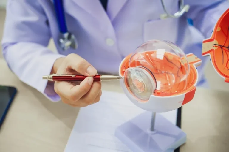

4 Techniques Used by Retina Specialists
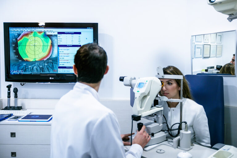
Retina specialists are ophthalmologists with advanced training in diseases of the retina and vitreous. After medical school and residency, they typically complete a fellowship focused on the back of the eye. They diagnose and manage conditions affecting this light-sensitive tissue, and these doctors use special instruments and techniques to treat a range of complex eye problems. Here are four techniques that retina specialists may use:
1. Pneumatic Retinopexy
This procedure addresses certain types of retinal detachments. The specialist injects a gas bubble into the eye, and the bubble then presses against the retina. This gentle pressure helps seal the retinal tear after the area is treated with cryotherapy or laser. Patients may need to maintain a specific head position for several days so the bubble remains in the correct place.
2. Focal Laser Therapy
Focal laser therapy is a precise treatment for specific retinal issues. It uses a targeted laser to treat small, localized areas. For conditions such as diabetic macular edema, the laser can seal leaking blood vessels, which helps reduce swelling. Specialists typically perform the procedure in an office setting.
This technique also helps create a barrier around a retinal tear, as the laser forms small burns around the tear, creating scar tissue. Scar tissue acts like a weld, and it secures the retina to the underlying tissue and prevents fluid from passing through the tear. This process helps stop the tear from progressing to a full retinal detachment.
3. Scleral Buckle
A scleral buckle is a surgical procedure that helps repair a retinal detachment. A retina specialist places a band, typically made of silicone, around the outside of the eye. After the procedure, the band gently presses the eye wall inward against the detached retina.
This indentation helps the retina reattach to the wall of the eye. In many cases, the buckle is typically left in place permanently. Specialists typically combine this procedure with other treatments, such as laser or cryotherapy, and it can seal any holes or tears in the retina.
By securing the retina in its proper position, the scleral buckle helps restore normal retinal function and preserves vision. While the band remains in place permanently, it is typically not noticeable and does not interfere with daily activities. Recovery from scleral buckle surgery may take several weeks, during which patients are advised to avoid strenuous activities and follow their surgeon’s instructions carefully.
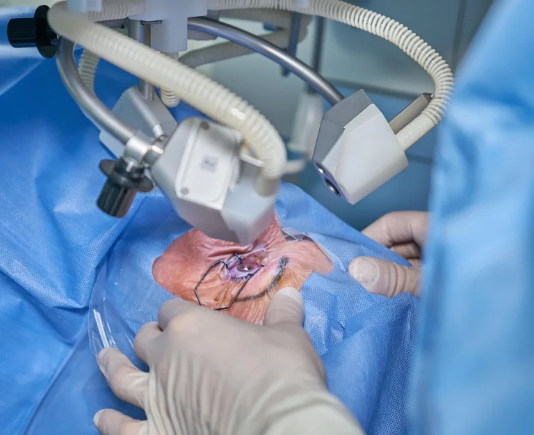
4. Surgical Vitrectomy
A surgical vitrectomy involves removing the vitreous gel from the middle of the eye. This procedure provides the surgeon with access to the retina. The surgeon makes small incisions in the white part of the eye, called the sclera, to introduce tiny instruments. They use these instruments to cut and remove the vitreous.
Once the vitreous is removed, the surgeon can address the underlying retinal problem. This may involve:
- Removing scar tissue
- Repairing retinal detachments
- Treating macular holes
At the end of the surgery, the eye is typically refilled with a saline solution, gas bubble, or silicone oil. This substance helps hold the retina in place while it heals. The choice of substance depends on the specific condition being treated.
Visit a Retina Specialist
Understanding these techniques offers insight into how retinal conditions are managed. A retina specialist can provide a thorough evaluation of your eye health. If you are experiencing changes in your vision, schedule an appointment with a qualified professional for an examination.